
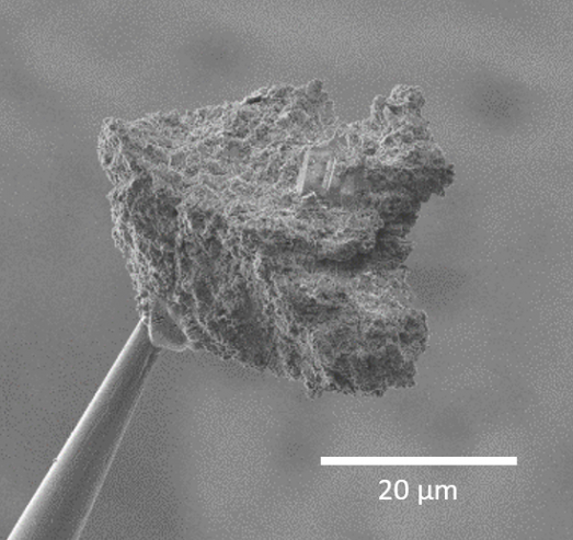
First IR and X-ray micro-tomography of asteroidal grains
How to measure and visualize in 3D the composition and structure of an extraterrestrial micrometric grain without destroying it? Researchers from the "Astrochemistry and Origin" team at IAS and the SOLEIL synchrotron, in collaboration with two Japanese laboratories, have demonstrated that this is possible on samples from the Itokawa asteroid collected by the Hayabusa spacecraft (JAXA), by coupling IR and X-ray micro-tomography.
The "Astrochemistry and Origins" team at IAS has specialized for many years in the study of small bodies of the Solar System (asteroids, comets, and interplanetary dust particles), with particular expertise in the infrared (IR) analysis of extraterrestrial grains in the laboratory. Recently, thanks to technological advances in IR detectors, a new barrier has been crossed, and IR hyperspectral imaging can now be performed in 3D at the micrometric scale.
X-ray micro-tomography is a 3D imaging technique that provides information on the physical properties of a sample (such as the shapes of various constituents, porosity, and 3D assembly). While X-ray micro-tomography is a well-established technique in numerous research fields (particularly in medicine), IR micro-tomography is a novel methodology. Recently implemented at the SOLEIL synchrotron through an international collaboration led by the IAS Astrochemistry and Origin team and SOLEIL's SMIS beamline, IR micro-tomography allows for probing the 3D composition of micrometric samples in a completely non-destructive manner. This technique is complementary to X-ray micro-tomography, as it is sensitive to the vibrational modes of chemical bonds within the molecules comprising the matter, providing insights into the 3D distribution of organic matter and minerals, as well as their local degree of hydration.
This coupling of IR and X-ray micro-tomography—a world first—was successfully carried out by researchers from the "Astrochemistry and Origin" team on six extraterrestrial grains and has just been published. The IR analysis was performed at SMIS-SOLEIL (France), while the X-ray analyses were conducted at SOLEIL's PSICHE beamline and beamline BL47XU at the Japanese SPring-8 synchrotron. These grains, ranging in diameter from 30 to 50 µm, have two distinct origins:
(i) Five grains were collected by the Hayabusa mission (JAXA) directly from the surface of the Itokawa asteroid.
(ii) One micrometric fragment of the Paris meteorite, one of the least altered carbonaceous chondrites present on Earth. This sample proved to be particularly interesting as it contains aliphatic organic matter.
Analysis of the Itokawa samples proved that combining IR and X-ray micro-tomography is viable for grains recovered from sample-return missions (Fig. 1). The combination of these techniques enabled the study of the mineral composition and 3D assembly of the asteroidal samples.
The study of the organic-rich Paris meteorite demonstrated that the heterogeneous 3D spatial distribution of aliphatic organic matter and its composition can be detected using non-destructive techniques, thereby avoiding any chemical or physical alteration of the grain (Fig. 2).
Recent sample-return missions Hayabusa2 (JAXA) and OSIRIS-REx (NASA), targeting the carbonaceous asteroids Ryugu and Bennu respectively, will yield only limited sample quantities (a few grams at most). Consequently, an analytical strategy must be devised that maximizes scientific return while minimizing sample loss. The combination of IR and X-ray micro-tomography can serve as the initial phase of a multi-analytical protocol, as it illuminates the 3D distribution of organic and mineral phases in extraterrestrial matter non-destructively.
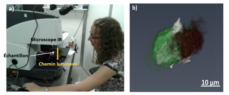
Figure 1: (a) The IR micro-tomography setup installed on the SMIS beamline at the SOLEIL synchrotron. (b) 3D micro-tomography reconstruction of the Itokawa particle RA-QD02-0214. Olivine (green) and pyroxene (brown) minerals are detected and clearly resolved using IR micro-tomography. The IR images are superimposed on X-ray micro-tomography data (in grey), which provide a high-resolution shape model of the particle.
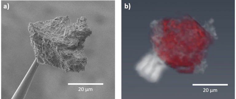
Figure 2: (a) Scanning electron microscope image of a grain from the Paris meteorite (b) 3D distribution of aliphatic organic matter obtained via IR micro-tomography (red), superimposed on the total 3D distribution of matter obtained via X-ray micro-tomography (grey).
References
Related articles:
– Z. Dionnet, R. Brunetto, A. Aléon-Toppani, S. Rubino, D. Baklouti, F. Borondics, A-C. Buellet, Z. Djouadi, A. King, T. Nakamura, A. Rotundi, C. Sandt, D. Troadec and A. Tsuchiyama, 2020. Combining IR and X-ray micro-tomography data sets: Application to Itokawa particles and to Paris meteorite. Meteoritics and Planetary Science 55, 1645-1664.
– A. Aléon-Toppani, R. Brunetto, J. Aléon, Z. Dionnet, S. Rubino, D. Levy, D. Troadec, F. Borondics and F. Brisset, 2020. A Preparation Sequence for Multi-Analysis of Micrometer-Sized Extraterrestrial Samples. Lunar and Planetary Science Conference, LPI Contribution No. 2326.
– R. Brunetto and C. Lantz, 2019. Laboratory perspectives on sample returns from Hayabusa2 and OSIRIS-REx. Nature Astronomy 3, 290–292.